

With summer in full swing, it is time to break out the sandals and open toed shoes. However, rising temperatures also mean your risk of developing a fungal infection is increased. Fungal infections of the hair, skin, and nail are caused by fungi called dermatophytes, in addition to molds and yeasts. Dermatophytes can be classified according to their natural habitat (zoophilic, anthropophilic, geophilic). For example, zoophilic dermatophytes infect animals, but can also transfer to humans through contact. Anthropophilic dermatophytes transfer from human to human, and geophilic dermatophytes dwell in soils. Dermatophyte infections can have different presentations depending on the causative pathogen and can be localized to specific areas of the body such as the scalp (Tinea capitis), nails (onychomycoses), and feet (Tinea pedis).
Without proper diagnosis and treatment, dermatophyte infections can lead to permanent damage of the hair, skin, and nails. Dermatophyte infections affect 20-25% of the global population1 and are particularly risky for the elderly and immunocompromised patients. Additionally, incorrect treatment can lead to worsening symptoms. Therefore, before starting an anti-fungal treatment, it is critical the true cause of the infection is accurately detected. The accurate detection of a fungus can be used to exclude other possible causes of a patient’s symptoms (psoriasis vulgaris, eczema, lichen planus, etc.).
What traditional methods are available to detect dermatophyte infections?
Dermatophyte infections are difficult to diagnose clinically as they can resemble other skin disorders. The identification of fungal pathogens is conventionally done with microscopy or fungal culturing. However, while a microscopic examination can give a direct positive or negative result as to whether a fungus is present but is limited in that it cannot identify the specific fungal species present. Species-specific identification is important as it can guide treatment duration, medication dosage, and allows for the possible determination of the source of an infection. For example, the detection of the fungal species Trichophyton mentagrophytes would point to a patient’s pet guinea pig as the possible carrier and cause of the infection. Subsequently the pet guinea pig could be treated appropriately, in addition to the human patient, thus preventing a recurrent infection.
Fungal culture is another common dermatophyte detection method and allows for the identification of the specific fungal species. However, fungi can take a long time to grow in culture, and therefore a patients’ results may only be obtained after about 4 to 6 weeks. Another limitation of culture is some fungal species do not grow in culture, and therefore a patient sample is at risk of being wrongly determined to be negative. A false-negative result leads to a patient not receiving proper therapy. Additionally, already initiated treatment (such as anti-fungal ointments) may prevent the fungal growth in culture and lead to false-negative results.
Molecular tests provide a fast and reliable detection of fungal infections
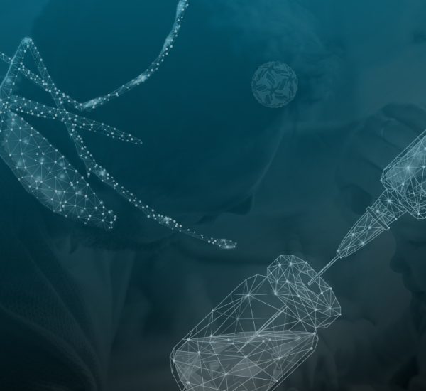
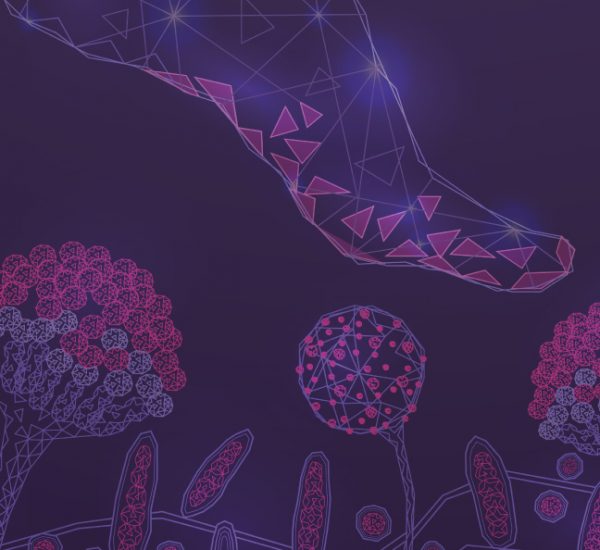
The limitations of conventional techniques for dermatophyte detection can be resolved through molecular diagnostics. Polymerase chain reaction (PCR)- based pathogen detection by DNA microarray allows for the direct, and specific identification of pathogens by means of their genetic material. Such molecular genetic detection shortens the diagnostic window from weeks to hours. The principle behind DNA microarray technology is that complementary strands of DNA will bind to each other. Once a patient sample is collected, DNA from the sample can be isolated. If a fungal pathogen is present on the patient sample, then the extract will contain a mixture of both human and fungal DNA. Using PCR, the DNA of a fungal pathogen (if present) is amplified and labelled with fluorescent probes. The PCR products can then be added to a BIOCHIP, a small glass chip containing a collection of microscopic DNA spots. If the complementary fungal DNA strand for a particular DNA spot is present in the PCR product, then the two strands will hybridize, i.e. bind to each other, and a fluorescent signal will be produced which can be measured. Therefore, the generation of a fluorescent signal will indicate that a fungus was present in the original patient sample.
The EUROArray Dermatomycosis solution utilizes the DNA microarray technique which provides direct detection and species differentiation of the most important dermatophytes in one PCR reaction. The entire procedure is easy to perform and takes just 3 hours from DNA isolation. The EUROArray Dermatomycosis detects 50 dermatophytes with 23 species-specific dermatophyte results in addition to six yeasts and molds. The direct detection allows for the accurate detection of dermatophytes that might be difficult to grow in culture, or in cases where a dermatophyte infection has already been treated. Further, species identification can point to the source of the infection, for example a pet in the case of a zoophilic dermatophyte. Molecular genetic methods such as the EUROArray Dermatomycosis enable fast and accurate pathogen-specific identification for the appropriate fungal infection treatment.
For more information on the EUROArray Dermatomycosis, and to see how this assay can be used by clinicians and labs, check out this previously recorded webinar from the EUROIMMUN Academy here.
References
- Havlickova B, Czaika VA, Friedrich M. Epidemiological trends in skin mycoses worldwide. Mycoses. 2008;51 Suppl 4:2-15.